


The ureteropelvic junction (UPJ) is the critical anatomic conduit where the funnel of the renal pelvis tapers into the proximal ureter. When this junction narrows or becomes mechanically obstructed, the normal propulsion of urine from the renal pelvis down to the bladder is blocked. This obstruction elevates retro-renal pressure, leading to progressive hydronephrosis (dilation of the kidney’s collecting system), chronic flank discomfort, recurrent urinary tract infections, secondary nephrolithiasis, and gradual loss of functional renal parenchyma.
Historically, surgical repair demanded open flank incisions that cut through major lateral abdominal muscle groups, necessitating prolonged hospital stays and extensive recovery periods. While traditional laparoscopy introduced a minimally invasive alternative, its rigid, non-articulating instruments proved technically demanding when executing delicate, watertight micro-suturing in a confined retroperitoneal corridor.
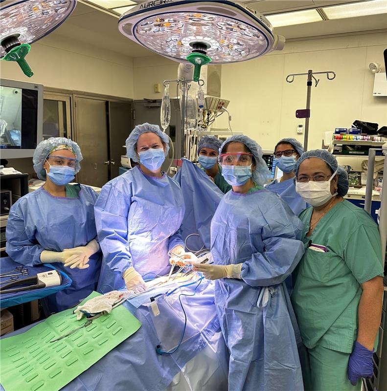
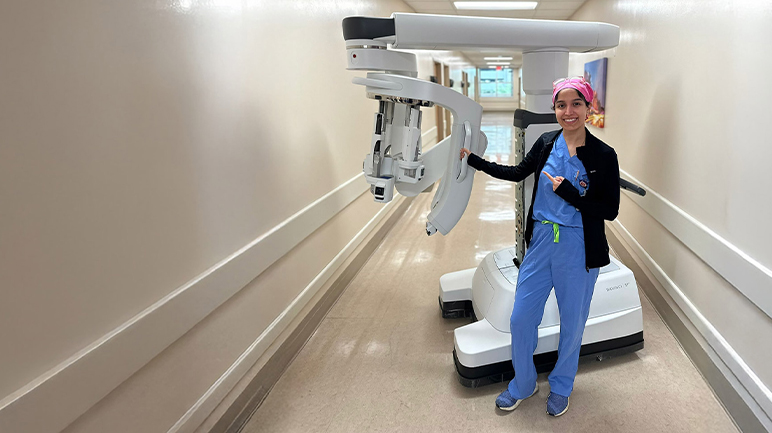
Today, Robotic-Assisted Pyeloplasty and Ureteral Reconstruction stands as the definitive gold standard for resolving UPJ obstructions and complex ureteral strictures. Practicing at Miami Robotic Surgery within the Comprehensive Urologic Surgery Institute in South Florida (miamiroboticsurgery.com), Dr. Shirin Razdan is a fellowship-trained urologic surgeon specializing in robotic urologic reconstruction. Having completed advanced fellowship training at the Icahn School of Medicine at Mount Sinai in New York—where she co-authored academic surgical literature on single-port and multi-port robotic reconstruction—Dr. Razdan utilizes multi-port da Vinci Xi and advanced Single Port (SP) platforms to reconstruct the upper urinary tract, restore anatomical flow, and protect long-term kidney function.
Etiology and Pathophysiology: Mechanisms of UPJ Obstruction
A UPJ obstruction can stem from intrinsic architectural abnormalities or extrinsic mechanical compression:
[UPJ OBSTRUCTION ETIOLOGY]
│
├─► 1. Intrinsic Stenosis / Aperistaltic Segment
│ └─► Congenital interruption of circular smooth muscle fibers or post-inflammatory scar tissue
│
└─► 2. Extrinsic Vascular Compression (Crossing Lower-Pole Vessels)
└─► Accessory renal artery or vein crosses the UPJ, kinking the funnel outflow tract
- Intrinsic Obstruction: Often a congenital defect where the normal circular smooth muscle fibers of the renal pelvis fail to transition into the ureter, leaving an aperistaltic, fibrotic segment. As urine pools, the segment cannot propagate normal peristalsis. It can also develop secondary to chronic inflammation from impacted kidney stones or prior endoscopic instrumentation.
- Extrinsic Crossing Vessels: In roughly 30% to 50% of adult presentation cases, an aberrant or accessory lower-pole renal artery or vein drapes directly across the anterior aspect of the ureteropelvic junction. As the pelvis distends with urine, the junction drapes tightly over this non-yielding vascular bundle, acting as a mechanical clamp.
The Gold Standard: Anderson-Hynes Dismembered Pyeloplasty
The most versatile, durable, and physiologically anatomical repair for UPJ obstruction is the dismembered Anderson-Hynes pyeloplasty. Robotic assistance provides high-definition 3D visualization, tremor filtration, and 7 degrees of freedom to execute each phase of this micro-reconstructive procedure:
[Console Mobilization & Vascular Mapping]
│
▼
[Complete Excision of Diseased UPJ Segment & Pelvic Reduction]
│
▼
[Transposition (Anterior Mobilization if Crossing Vessel Present)]
│
▼
[Lateral Ureteral Spatulation (1.5–2 cm) for Broad Funnel]
│
▼
[Endo-Wristed Running Watertight Anastomosis over Double-J Stent]
- Retroperitoneal Mobilization & Vascular Dissection: The colon is carefully mobilized along the line of Toldt to reveal the retroperitoneal workspace. The dilated renal pelvis and upper ureter are isolated. If a lower-pole crossing vessel is identified, the pelvic-ureteral junction is mobilized so the repair can be transposed anterior to the blood vessels, relieving extrinsic mechanical compression.
- Dismemberment & Excision: The diseased, aperistaltic, or fibrotic junction is excised completely. If chronic hydronephrosis has ballooned the renal pelvis into a large, non-draining sac, a reduction pyeloplasty is performed—trimming redundant pelvic tissue to optimize future fluid dynamics.
- Ureteral Spatulation: The healthy proximal ureter is spatulated (incised longitudinally along its lateral border) for 1.5 to 2 centimeters. This critical step converts a narrow circular tube into a wide, open trough, creating a broad funnel that prevents future stricture recurrence.
- Endo-Wristed Running Anastomosis: Utilizing micro-sutures, the spatulated ureter is anastomosed to the lowest, most dependent portion of the newly fashioned renal pelvis. The wristed instruments allow precise, tension-free suture placement along both the posterior and anterior plates.
- Internal Stenting: A Double-J ureteral stent is placed across the reconstruction to divert urine, protect the healing tissues, and keep the anastomosis decompressed. The stent is removed in the office 4 to 6 weeks later.
Beyond the UPJ: Complex Ureteral Reconstructions
In addition to primary UPJ obstruction, Dr. Shirin Razdan manages complex proximal and mid-ureteral strictures resulting from previous stone impaction, radiation therapy, endometriosis, or surgical trauma:
┌──► 1. Ureteroureterostomy (Short focal mid-ureteral strictures)
│ ──► Excision of fibrotic segment followed by end-to-end spatulated re-anastomosis
│
[COMPLEX RECONSTRUCTIVE SPECTRUM] ┼──► 2. Buccal Mucosa Onlay Graft (BMG) Ureteroplasty
│ ──► Oral mucosal graft patch for long, non-transectable strictures
│
└──► 3. Ureteral Reimplantation with Boari Flap / Psoas Hitch
──► Mobilizing bladder upward to bridge distal ureteral defects
- Buccal Mucosa Onlay Graft (BMG) Ureteroplasty: For long, dense strictures ($> 2\text{ cm}$) where direct end-to-end re-approximation would cause excessive tension, Dr. Razdan utilizes an autologous buccal mucosa graft harvested from the patient’s inner cheek. The ureter is incised longitudinally across the stricture, and the oral graft is sutured into place as a patch graft to widen the lumen, often wrapped in an omental flap to promote vascular ingrowth.
- Ureteral Reimplantation (Ureteroneocystostomy): For lower ureteral strictures, the healthy proximal ureter is directly re-implanted into the bladder dome with an anti-reflux tunnel, frequently supported by a Psoas Hitch (anchoring the bladder to the psoas minor tendon) or a tubularized Boari Flap of bladder wall to bridge long defects without tension.
Surgical Platforms: da Vinci Multi-Port (Xi) vs. Single Port (SP)
Dr. Razdan tailors the robotic platform to the patient’s surgical history, body habitus, and stricture complexity:
| Reconstructive Feature | Traditional Laparoscopy | Multi-Port Robotics (da Vinci Xi) | Single Port Robotics (da Vinci SP) |
| Incision Profile | 3 to 4 scattered keyhole trocars | 3 to 4 ports across abdomen | 1 single 2.5 cm incision (umbilical) |
| Instrument Articulation | Rigid straight instruments (no wrist) | 7 degrees of freedom wristed micro-shears | Double-jointed (“elbow & wrist”) instruments |
| Suturing Precision | Technically challenging in tight spaces | High precision; rapid running sutures | High precision in deep, narrow corridors |
| Bowel Mobilization | Requires transperitoneal bowel retraction | Standard transperitoneal or retroperitoneal | Direct retroperitoneal access (no bowel entry) |
| Post-Operative Pain | Mild to moderate abdominal soreness | Mild; well-managed with oral analgesics | Minimal; muscle-sparing narcotic-free recovery |
| Durable Patency Rate | 88% to 92% | 95% to 98% long-term success | 95% to 98% long-term success |
Preoperative Diagnostic Roadmap and Functional Verification
Accurate structural and functional localization is mandatory to ensure that the patient’s symptoms correlate directly with physiological obstruction:
- MAG3 Diuretic Renal Scintigraphy: Confirms delayed radiotracer clearance with an obstructive clearance half-time ($T_{1/2} > 20\text{ minutes}$) following intravenous administration of Lasix, while quantifying differential split renal function.
- High-Resolution CT Angiography / CT Urogram: Provides thin-slice mapping of the renal parenchyma, collecting system, and vascular tree, confirming or excluding lower-pole crossing renal vessels.
- Postoperative Functional Verification: Several months after stent removal, a follow-up ultrasound and repeat MAG3 scan verify resolution of hydronephrosis and confirm rapid, unobstructed drainage ($T_{1/2} < 10\text{ minutes}$) with stable renal function.
Conclusion
UPJ obstruction and ureteral strictures do not require muscle-splitting open operations or permanent nephrostomy tubes. Through robotic-assisted pyeloplasty and advanced ureteral reconstruction—utilizing multi-port and single-port platforms—Dr. Shirin Razdan provides long-term anatomical resolution, preserving renal function and helping patients return to their daily lives with minimal recovery downtime.
- Surgeon: Dr. Shirin Razdan, MD
- Specialty: Fellowship-Trained Robotic Urologic Surgeon & Reconstructive Urologist
- Practice: Miami Robotic Surgery / Comprehensive Urologic Surgery Institute
- Office Location: 3650 NW 82nd Avenue, Suite 502, Doral/Miami, FL 33166
- Official Website: miamiroboticsurgery.com
- Direct Consultations & Inquiries: (305) 468-3314