



The widespread adoption of cross-sectional abdominal imaging has made the incidental detection of a small renal mass (SRM)—defined as a solid or complex cystic enhancing renal lesion measuring 4cm (clinical stage cT1a)—one of the most common encounters in contemporary urology. Most of these lesions cause no symptoms, such as flank pain or hematuria, and are identified during evaluations for unrelated abdominal issues.
A generation ago, discovering a solid renal mass triggered immediate, radical extirpation: total removal of the entire kidney and its surrounding adrenal gland. Modern urologic oncology operates under a fundamentally different paradigm. Surgical and pathological series demonstrate that 20% to 30% of solid small renal masses under 4 cm are completely benign (such as oncocytomas or lipid-poor angiomyolipomas). Among the remaining 70% to 80% that represent renal cell carcinoma (RCC), the vast majority are low-grade tumors exhibiting indolent biological behavior, with annual linear growth rates averaging only 0.2 to 0.3 cm per year and low 3-year metastatic rates ($\sim 1\%\text{–}2\%$).
Navigating a small kidney mass requires choosing between three distinct pathways: Active Surveillance (AS), Thermal Ablation, or Nephron-Sparing Robotic Partial Nephrectomy (PN).
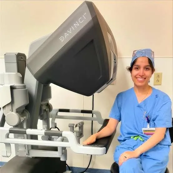
Practicing in Miami, Florida, Dr. Shirin Razdan, MD is a dual-fellowship trained, board-certified urologist specializing in advanced robotic urologic oncology and complex endourology. Having completed intensive robotic surgery and oncology fellowships at the Icahn School of Medicine at Mount Sinai in New York City, Dr. Razdan directs robotic surgery at the Comprehensive Urological Surgery Institute in Miami. Utilizing high-resolution cross-sectional imaging, R.E.N.A.L. nephrometry scoring, and selective percutaneous renal biopsies, Dr. Razdan tailors the management strategy to the biological characteristics of the tumor and the functional health of the patient.
Evaluating the Three Management Pathways
Each clinical approach is designed for specific patient populations, anatomical complexities, and biological profiles:
1. Robotic Partial Nephrectomy (The Curative Gold Standard)
The American Urological Association (AUA) guidelines state that surgeons should prioritize partial nephrectomy over radical nephrectomy whenever intervention is indicated for a cT1a mass.
- The Technique: Dr. Razdan utilizes multi-port or single-port robotic platforms to precisely excise the tumor with a thin margin of normal parenchyma, followed by reconstruction of the renal defect and collecting system under minimal ischemia time.
- The Strengths: Delivers the highest long-term oncologic cure rates (local control $> 95\%\text{–}98\%$), provides a full physical specimen for precise pathological grading and margin evaluation, and preserves maximal nephron mass to protect long-term cardiovascular and renal health.
- Best Suited For: Young, healthy, surgically fit patients; tumors between 2 and 4 cm; complex central or hilar anatomy; or patients seeking definitive histopathological confirmation and cure.
2. Percutaneous Thermal Ablation (Cryoablation & Microwave/RFA)
A minimally invasive approach that destroys malignant tissue in situ without surgical excision.
- The Technique: Guided by real-time CT or ultrasound, an interventional radiologist or urologist inserts specialized probes through the skin directly into the tumor center. Freezing temperatures (cryoablation down to $-40^\circ\text{C}$) or extreme heat (microwave/radiofrequency ablation $> 60^\circ\text{C}$) induce cellular rupture and microvascular thrombosis.
- The Strengths: Performed on an outpatient basis under conscious sedation or light anesthesia with minimal systemic physiological stress and zero surgical incisions.
- The Trade-Offs: Carries a slightly higher rate of local persistence or recurrence compared to surgical resection (often requiring secondary ablation or follow-up imaging). It is less effective for tumors >3cm or central lesions situated adjacent to the renal hilum or ureter (due to the “heat-sink” effect of large blood vessels or thermal injury risks to the renal pelvis).
- Best Suited For: Frail or elderly patients with significant cardiopulmonary comorbidities who carry high general anesthesia risks, especially with exophytic tumors <3cm located along the peripheral renal cortex.
3. Active Surveillance (Safe Delayed Intervention)
Active surveillance involves structured serial cross-sectional imaging to monitor the tumor’s growth kinetics, reserving active intervention for tumors that demonstrate progressive growth or concerning features.
- The Protocol: Abdominal CT, MRI, or renal ultrasound performed every 3 to 6 months for the first year to calculate the linear growth rate, followed by semi-annual or annual scans.
- The Safety Profile: Extensive prospective registry data confirms that delaying intervention does not compromise overall survival or curability for indolent small masses. If a tumor grows rapidly (>0.5cm/year) or reaches 3 to 4 cm, delayed partial nephrectomy or ablation is performed without oncologic penalty.
- Best Suited For: Tumors <2cm; patients with advanced age or competing medical illnesses; individuals with severe baseline chronic kidney disease (CKD); or patients prioritizing the avoidance of immediate procedures.
The Decision Matrix: Comparing Management Options
| Decision Factor | Active Surveillance | Thermal Ablation (Cryo/RFA) | Robotic Partial Nephrectomy |
| Invasiveness | None (Serial cross-sectional imaging) | Minimally invasive percutaneous needles | Minimally invasive keyhole robotic surgery |
| Anesthesia Required | None | Local sedation or light general anesthesia | Full general endotracheal anesthesia |
| Histological Specimen | Only if percutaneous biopsy performed | Core needle biopsy prior to probe activation | Complete intact specimen; verified margins |
| Local Tumor Control | N/A (Deferred treatment) | 88% to 92% primary success (repeatable) | 95% to 98%+ definitive local control |
| Complication Risk | Psychological anxiety; rare progression | Low (Bleeding, adjacent thermal injury) | Low-to-moderate (Bleeding, urine leak) |
| Ideal Tumor Size | Best for <2cm | Best for exophytic tumors <3cm | Up to 4 cm (and select larger cT1b lesions) |
Frequently Asked Questions (FAQ)
Should I have a percutaneous biopsy of my small kidney mass?
AUA guidelines recommend a renal mass biopsy (RMB) whenever the result would alter the clinical plan. Biopsy is particularly valuable prior to thermal ablation (to confirm malignancy and guide follow-up) or when deciding between surveillance and surgery in older adults. A biopsy is generally omitted in young, healthy patients who prefer definitive surgical removal regardless of whether the mass is low-grade cancer or an indeterminate neoplasm.
Will I lose my entire kidney if I undergo surgery for a small mass?
In the hands of an experienced robotic urologic oncologist, complete kidney removal (radical nephrectomy) is rarely necessary for an uncomplicated mass under 4 cm. Robotic partial nephrectomy excises only the tumor along with a thin rim of tissue, preserving the healthy renal cortex to protect total filtration function.
What specific changes trigger an exit from active surveillance to surgery?
Treatment is typically recommended if the tumor demonstrates a rapid linear growth rate (>0.5cm/year), reaches an absolute size threshold of 3 to 4 cm, shows evidence of infiltrative growth on contrast MRI, or if the patient develops persistent hematuria or requests treatment.
Conclusion
A small kidney mass under 4 cm is an actionable finding, but it rarely represents an immediate oncologic emergency. Because many small renal lesions are benign or slow-growing, reflexively removing the kidney is an outdated approach. By weighing individual tumor size, anatomic complexity, nephron reserve, and overall health status, patients can choose safely among structured surveillance, image-guided ablation, or robotic partial nephrectomy. Consulting with a dedicated robotic urologic surgeon ensures an individualized, evidence-based management strategy.