


Unlike many common solid malignancies, such as breast, colon, or lung cancer, renal cell carcinoma (RCC) has no recommended routine screening blood test or universal screening protocol. Instead, more than 70% of modern kidney tumors are discovered incidentally during cross-sectional imaging performed for unrelated abdominal or pelvic symptoms, such as gastrointestinal distress, flank pain, or trauma workups.
Once an unexpected renal mass is visualized on a routine screening ultrasound or non-contrast scan, the clinical objective shifts immediately to definitive characterization. A urologic oncologist must answer three critical clinical questions: Is the lesion solid or cystic? Does it exhibit internal vascular enhancement consistent with malignancy? Is surgical extirpation, percutaneous ablation, or active surveillance the appropriate management path?
Answering these questions relies on three core diagnostic modalities: Multiphasic Computed Tomography (CT), Multiparametric Magnetic Resonance Imaging (MRI), and selective Renal Mass Biopsy (RMB).
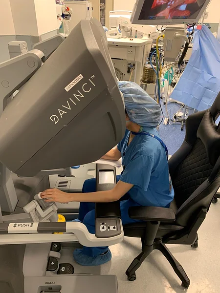
Practicing in Miami, Florida, Dr. Shirin Razdan, MD is a dual-fellowship trained, board-certified urologist specializing in advanced robotic urologic oncology and complex endourology. Having completed intensive robotic surgery and oncology fellowships at the Icahn School of Medicine at Mount Sinai in New York City, Dr. Razdan serves as the Director of the Robotic Surgery Department at the Comprehensive Urological Surgery Institute in Miami. Applying specialized imaging protocols, objective anatomical scoring metrics, and targeted percutaneous biopsies, Dr. Razdan guides patients through an evidence-based diagnostic workup tailored to preserve renal function and optimize oncologic outcomes.
1. Multiphasic Contrast-Enhanced CT: The Imaging Workhorse
A standard non-contrast CT scan or a single-phase abdominal scan with oral contrast is insufficient to evaluate a suspected renal neoplasm. The gold-standard radiographic evaluation for renal tumors is a dedicated multiphasic renal protocol CT, which captures images across three distinct contrast phases:
- Unenhanced (Pre-Contrast) Phase: Establishes the baseline attenuation of the mass measured in Hounsfield Units (HU). This phase distinguishes between calcifications, high-density benign proteinaceous cysts, and microscopic fat (which confirms a benign angiomyolipoma).
- Corticomedullary Phase (25 to 70 seconds post-injection): Highlights maximal contrast enhancement within the renal cortex. Hypervascular clear cell renal cell carcinomas light up intensely during this early phase, allowing precise delineation of tumor borders relative to the cortical parenchyma.
- Nephrographic Phase (80 to 180 seconds post-injection): The contrast agent distributes uniformly throughout the renal medullary parenchyma. This phase offers the highest sensitivity for detecting subtle, hypovascular tumors (such as papillary RCC or chromophobe RCC) and identifies small endophytic lesions buried within the kidney.
- Excretory / Delayed Phase (5 to 10 minutes post-injection): Contrast opacifies the renal calyces, renal pelvis, and ureter, demonstrating whether the tumor compresses or directly invades the urinary collecting system—a vital consideration when planning robotic partial nephrectomy.
The defining radiographic hallmark of kidney cancer is measurable contrast enhancement. If a solid mass demonstrates an increase of 15 to 20 Hounsfield Units or greater between the pre-contrast and post-contrast phases, it is treated as a malignant renal neoplasm until proven otherwise.
2. Multiparametric Renal MRI: When and Why It Is Selected
While multiphasic CT is the most common diagnostic tool, multiparametric MRI (mpMRI) provides superior soft-tissue contrast and cellular characterization without ionizing radiation.
MRI is specifically indicated in three clinical settings:
- Severe Renal Insufficiency or Iodine Allergy: Patients with chronic kidney disease (CKD) or anaphylactic allergies to iodinated CT contrast agents can undergo contrast-enhanced MRI utilizing specific gadolinium-based agents, or non-contrast diffusion-weighted MRI protocols.
- Evaluating Vascular Tumor Thrombus: In locally advanced tumors, cancer cells can invade the renal vein and propagate into the inferior vena cava (IVC). MRI provides superior evaluation of the superior extent of the vascular thrombus and assesses whether the tumor has physically invaded the vena caval endothelial wall, which dictates surgical vascular clamping strategies.
- Characterizing Complex Cystic Masses (The Bosniak Classification): Fluid-filled kidney cysts range from harmless simple cysts to complex cystic renal carcinomas. MRI is exceptionally sensitive for evaluating internal septations, wall thickness, and subtle mural nodules under the Bosniak Classification System (Categories I through IV), guiding decisions between observation (Bosniak II/IIF) and robotic excision (Bosniak III/IV).

3. Renal Mass Biopsy (RMB): Role, Indications, and Myths
Historically, urologists avoided percutaneous needle biopsies of kidney masses due to fears of “tumor tract seeding” (accidentally dragging cancer cells along the needle path) and bleeding. Modern biopsy needles utilize coaxial cutting sheaths that shield the needle path, reducing the statistical risk of tumor seeding to negligible rates ($< 0.01\%$).
According to American Urological Association (AUA) guidelines, a renal mass biopsy is not universally required for every surgically fit patient with a classic, enhancing solid mass who is already committed to surgical removal. However, biopsy is strongly recommended when its findings will directly alter clinical management:
- Prior to Thermal Ablation or SBRT: Because freezing (cryoablation), heating (radiofrequency), or radiation (SBRT) destroys tissue without producing a surgical specimen, a core needle biopsy is mandatory before treatment to confirm malignancy and define tumor histology.
- Candidates for Active Surveillance: When considering observation for a small renal mass in an older or medically frail adult, biopsy confirms whether the tumor is an indolent, low-grade lesion or an aggressive Grade 4 variant requiring active treatment.
- Suspected Metastasis, Lymphoma, or Benign Neoplasm: Biopsy is critical when clinical findings suggest an inflammatory abscess, renal lymphoma, or metastatic spread from another primary cancer (such as lung, breast, or melanoma), where kidney surgery would be inappropriate. It also definitively identifies benign oncocytomas and lipid-poor angiomyolipomas.
Diagnostic Comparison: Evaluating Modalities
| Diagnostic Modality | Primary Diagnostic Role | Key Strengths | Limitations |
| Multiphasic Renal CT | Initial evaluation and surgical planning | Rapid, standardized, high-spatial resolution; quantifies Hounsfield Units | Involves ionizing radiation and iodinated contrast load |
| Multiparametric MRI | Indeterminate masses, cysts, and IVC thrombi | Exceptional soft-tissue contrast; maps vascular extension without CT contrast | Longer scan time, higher cost; claustrophobia considerations |
| Renal Mass Biopsy | Histological typing and nuclear grading | Confirms cancer vs. benign; guides ablation and surveillance | Invasive core needle; 10%–15% rate of non-diagnostic sampling |
Frequently Asked Questions (FAQ)
Can a blood or urine test detect kidney cancer?
No. There are currently no validated blood or urine biomarkers approved for the initial detection of renal cell carcinoma. Routine laboratory work—such as serum creatinine, estimated GFR, urinalysis (checking for microscopic hematuria), and complete blood counts—is vital for evaluating baseline kidney filtration and systemic health, but imaging remains mandatory for diagnosis.
What percentage of small solid kidney tumors turn out to be benign?
Approximately 15% to 20% of surgically removed solid renal masses under 4 cm (cT1a) are ultimately diagnosed on final surgical pathology as completely benign entities, most commonly oncocytomas or lipid-poor angiomyolipomas. This reality highlights the growing role of selective pre-treatment core needle biopsies for small masses.
What is a R.E.N.A.L. Nephrometry Score?
The R.E.N.A.L. nephrometry score is a standardized anatomical grading system that urologists calculate from your CT or MRI. It scores five structural characteristics: Radius (tumor size), Exophytic/endophytic properties (how deeply it sits inside the kidney), Nearness to the collecting system, Anterior/posterior orientation, and Location relative to the polar lines. This score quantifies surgical complexity and helps Dr. Razdan determine whether robotic partial nephrectomy is technically safe and feasible.
Conclusion
Accurate diagnosis of kidney cancer requires a structured, multi-step assessment that moves beyond non-specific ultrasound findings. By leveraging multiphasic contrast-enhanced CT protocols, utilizing multiparametric MRI for vascular and cystic delineation, and deploying targeted core biopsies when histological clarity alters care, patients receive an objective assessment of their tumor. Through detailed anatomical scoring and minimally invasive robotic expertise, Dr. Shirin Razdan ensures that diagnostic clarity translates into a personalized, nephron-sparing treatment strategy.